

The intersection of herpetology and neuroscience has yielded remarkable advances in our understanding of the human brain. Snakes, with their unique neurological adaptations and specialized sensory systems, have become invaluable subjects in cutting-edge neurological research. From their specialized venom compounds that target specific neural pathways to their remarkable sensory organs that detect infrared radiation and chemical signals, snakes offer neuroscientists a treasure trove of evolutionary innovations. This article explores the fascinating ways researchers are utilizing snake biology to develop new treatments for neurological disorders, uncover fundamental principles of neural processing, and inspire biomimetic technologies that could revolutionize medicine.
The Therapeutic Potential of Snake Venoms
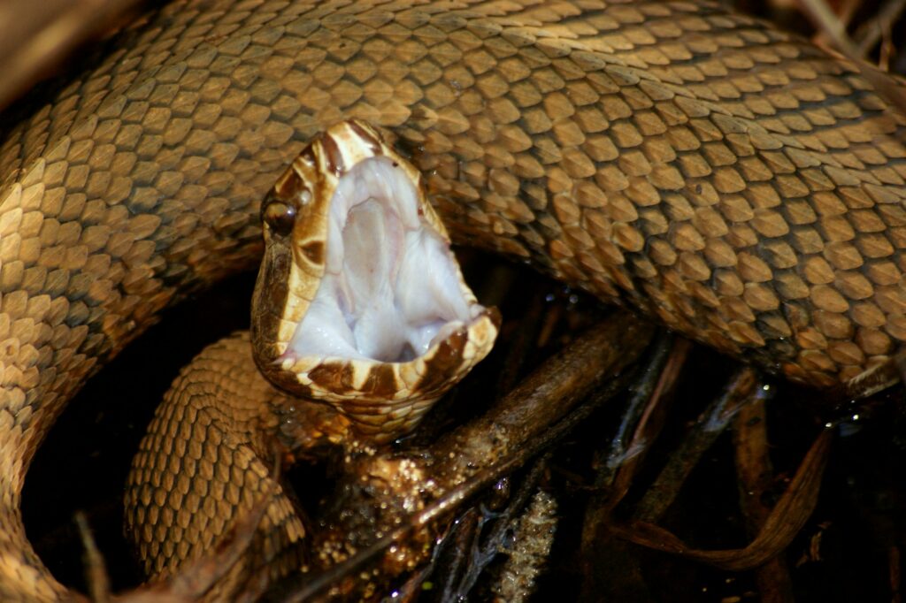
Snake venoms contain a complex cocktail of bioactive compounds that have evolved to target specific neurological systems, making them invaluable tools for drug discovery. Researchers have identified numerous peptides and proteins in snake venoms that interact with ion channels, neurotransmitter receptors, and other crucial components of the nervous system with remarkable precision. One notable success story is the development of captopril, a medication for hypertension derived from peptides found in the venom of the Brazilian pit viper (Bothrops jararaca). More recently, scientists have isolated compounds from cobra venom that show promise in treating Alzheimer’s disease by inhibiting the formation of amyloid plaques. These naturally occurring compounds often possess specificity and potency far exceeding synthetic alternatives, providing researchers with molecular scaffolds that can be modified to develop targeted neurological treatments.
Studying Pain Pathways Through Venom Research

Snake venoms have become powerful tools for understanding pain pathways in the human nervous system. Many venom components specifically target ion channels involved in pain transmission, such as voltage-gated sodium channels and acid-sensing ion channels. By studying how these toxins interact with their targets, researchers can elucidate the structure and function of these channels and develop new analgesic drugs. The venom of the Texas coral snake (Micrurus tener), for instance, contains toxins that block certain pain-signaling pathways while leaving other neural functions intact. This specificity provides neuroscientists with precision tools to study discrete components of the pain response. Such research has already led to the development of novel pain medications that avoid the addiction risks and side effects associated with opioids by targeting different neural mechanisms altogether.
Infrared Sensing and Neural Processing

Pit vipers, pythons, and boas possess specialized sensory organs known as pit organs that allow them to detect infrared radiation with extraordinary sensitivity. These organs can detect temperature differences as small as 0.003°C, enabling the snakes to locate warm-blooded prey in complete darkness. Neuroscientists are studying these remarkable organs to understand how thermal information is processed and integrated with visual data in the brain. The trigeminal nerve pathways that transmit signals from pit organs to the snake’s optic tectum represent a unique example of multimodal sensory integration. Recent research using functional imaging techniques has revealed that pit vipers essentially “see” in infrared, with thermal information mapped alongside visual data in their brain’s spatial representation. These findings have profound implications for understanding how different sensory modalities can be integrated in the brain and may inspire new approaches to sensory augmentation for humans with impaired vision.
Venom Production as a Model for Neurosecretory Processes

The specialized glands that produce snake venom represent exceptional models for studying neurosecretory processes. These highly specialized secretory systems undergo dramatic ultrastructural changes during venom production, with complex transcriptional regulation controlled by neuronal signals. Researchers have discovered that many of the cellular mechanisms involved in venom production are similar to those used by neurons to release neurotransmitters. The venom gland epithelial cells of snakes like the Eastern diamondback rattlesnake (Crotalus adamanteus) express many proteins typically associated with neuronal secretion. By studying the regulatory processes that control venom production, scientists gain insights into how neurons regulate the synthesis and release of signaling molecules. These discoveries have applications for understanding neurodegenerative diseases characterized by protein misfolding and secretory dysfunction, such as Parkinson’s disease.
Neuroanatomical Adaptations in Snakes

The extreme body elongation of snakes has resulted in fascinating neuroanatomical adaptations that provide unique research opportunities for neuroscientists. Unlike most vertebrates, snakes have undergone dramatic transformations in their neural architecture, including the reduction or loss of limb-related neural circuits and the expansion of trunk-related sensory and motor systems. Researchers have discovered that the somatosensory cortex of snakes shows specialized organization reflecting their limbless body plan. Studies of Burmese pythons (Python bivittatus) have revealed that the neural control of their synchronized, wave-like muscular contractions during locomotion involves specialized central pattern generators in the spinal cord. These adaptations provide models for understanding neural plasticity and reorganization following injury or evolutionary change. By examining how snake brains have evolved to control their unique body form, researchers gain insights into fundamental principles of neural organization that could inform rehabilitation strategies for patients with spinal cord injuries.
Vomeronasal System and Chemical Sensing

Snakes possess one of the most sophisticated chemical sensing systems in the animal kingdom, centered around the vomeronasal (Jacobson’s) organ and their iconic forked tongues. This system allows snakes to create a three-dimensional chemical map of their environment with remarkable sensitivity. Neuroscientists study this system to understand how chemical information is processed and integrated in the brain. Recent research using advanced imaging techniques has revealed that the snake’s vomeronasal system involves specialized neural circuits that process chemical signals differently from the main olfactory system. The forked tongue of snakes like the garter snake (Thamnophis sirtalis) acts as a stereochemical sampler, with each tine delivering samples to separate compartments of the vomeronasal organ. This sophisticated system provides insights into how spatial information can be encoded through chemical sensing, with potential applications for developing advanced artificial sensing technologies for detecting environmental toxins or diagnosing diseases through chemical biomarkers.
Neural Control of Extreme Feeding Adaptations

Many snake species have evolved extraordinary feeding adaptations, including the ability to consume prey much larger than their head diameter. These adaptations require sophisticated neural control systems to coordinate the complex muscular activities involved in prey capture and swallowing. Neuroscientists study these mechanisms to understand neural control of coordinated muscular movements. The feeding process in snakes like the king cobra (Ophiophagus hannah) involves independent movement of the left and right sides of the jaw, requiring precise neural coordination. Research using electromyography has revealed that the swallowing process is controlled by specialized neural circuits in the brainstem that generate rhythmic muscular contractions. Studying these neural control systems provides insights into the coordination of complex motor sequences and has applications for understanding and treating swallowing disorders in humans, which affect millions of patients following stroke or neurodegenerative disease.
Biomimetic Neural Interfaces Inspired by Snake Sensory Systems

The remarkable sensory adaptations of snakes have inspired innovative biomimetic technologies for neural interfaces and prosthetic devices. Engineers have developed infrared sensors based on the architecture of snake pit organs that could be integrated into neural prosthetics for the visually impaired. These bio-inspired sensors mimic the membrane structure and neural processing of pit organs to achieve sensitivity that far exceeds conventional infrared detectors. Similarly, the chemical sensing capabilities of the vomeronasal system have inspired the development of electronic “tongues” for medical diagnostics. Researchers at institutions like Stanford University have created flexible electronic interfaces that mimic the snake’s ability to detect subtle chemical signals. These biomimetic approaches demonstrate how understanding snake sensory systems can lead to technological innovations with direct applications in neurological rehabilitation and assistive technologies for patients with sensory impairments.
Unraveling Neural Regeneration Through Snake Research

Certain snake species demonstrate remarkable neural regenerative capabilities that provide insights into potential therapies for traumatic brain and spinal cord injuries. Unlike mammals, many reptiles, including snakes, show significant capacity for neural regeneration following injury. Studies on species like the Texas rat snake (Elaphe obsoleta) have revealed molecular pathways that permit neural regrowth that would be inhibited in mammals. Researchers have identified specific growth factors and cellular mechanisms that allow snake neurons to regrow axons and reestablish functional connections after damage. Comparative studies between mammalian and reptilian neural regeneration have highlighted key differences in inflammation responses and glial scar formation that may inform therapeutic approaches. By identifying the molecular switches that permit neural regeneration in snakes, researchers hope to develop interventions that could activate similar regenerative pathways in human patients with traumatic brain injuries or neurodegenerative conditions.
Snake Neurons as Models for Neurodegeneration Research
The extraordinary longevity of many snake species makes them valuable models for studying neurodegenerative processes and potential protective mechanisms. Some species, like the ball python (Python regius), can live for over 30 years, providing opportunities to study how neurons maintain functionality over extended periods. Researchers have discovered that snake neurons possess unique mechanisms for protein quality control and stress resistance that may protect against age-related neurodegeneration. Studies of protein aggregation in aging snake brains have revealed protective mechanisms that prevent the formation of amyloid plaques similar to those seen in Alzheimer’s disease. Additionally, the seasonal changes in brain structure and function that occur during hibernation in species like the timber rattlesnake (Crotalus horridus) provide insights into neural plasticity and neuroprotection during metabolic stress. These natural adaptations could inspire novel therapeutic approaches for neurodegenerative conditions in humans by mimicking the protective mechanisms observed in snake neurons.
Neurotoxins as Tools for Studying Synaptic Transmission

Snake neurotoxins have become indispensable tools for studying the molecular mechanisms of synaptic transmission in the nervous system. Toxins like α-bungarotoxin from the many-banded krait (Bungarus multicinctus) bind with extraordinary specificity to acetylcholine receptors, allowing researchers to visualize and quantify these receptors in neuronal membranes. Other neurotoxins selectively block specific ion channels or receptors, enabling researchers to dissect the contributions of different molecular components to neural signaling. The three-finger toxins found in cobra venoms have been particularly valuable for mapping the structure and function of nicotinic acetylcholine receptors involved in neuromuscular transmission. By using these toxins as molecular probes, researchers have made fundamental discoveries about synaptic plasticity and neurotransmitter receptor functioning. These findings have direct applications for understanding neurological disorders characterized by synaptic dysfunction, such as myasthenia gravis, certain forms of epilepsy, and various neurodegenerative conditions.
Ethical Considerations and Future Directions

As with all animal research, the use of snakes in neurological studies raises important ethical considerations that researchers must carefully address. The scientific community has developed strict guidelines for the humane treatment of reptiles in laboratory settings, emphasizing the principles of replacement, reduction, and refinement. Advanced techniques like non-invasive imaging, micro-sampling of venom, and computational modeling have reduced the need for invasive procedures in many research contexts. Looking toward the future, emerging technologies like organoids derived from snake tissues and advanced computational models may further reduce the need for live animal studies while still providing valuable insights. The continued development of interdisciplinary collaboration between herpetologists, neuroscientists, and bioethicists will be essential to ensure that snake-based neurological research proceeds ethically and sustainably. This balanced approach will maximize the scientific and medical benefits of this research while respecting the welfare of these remarkable creatures that have so much to teach us about the brain.
In conclusion, snakes represent an extraordinary and often overlooked resource for neurological research, offering unique adaptations that illuminate fundamental principles of neural function and inspire innovative therapeutic approaches. From venom compounds that target specific neural pathways with remarkable precision to sensory systems that detect stimuli beyond human perception, snake biology continues to drive advances in neuroscience. As technology evolves and interdisciplinary collaboration expands, the contribution of snake-based research to addressing neurological disorders is likely to grow substantially. By studying these remarkable reptiles with both scientific rigor and ethical consideration, researchers are uncovering insights that may ultimately transform our understanding of the human brain and our approach to treating its disorders.


